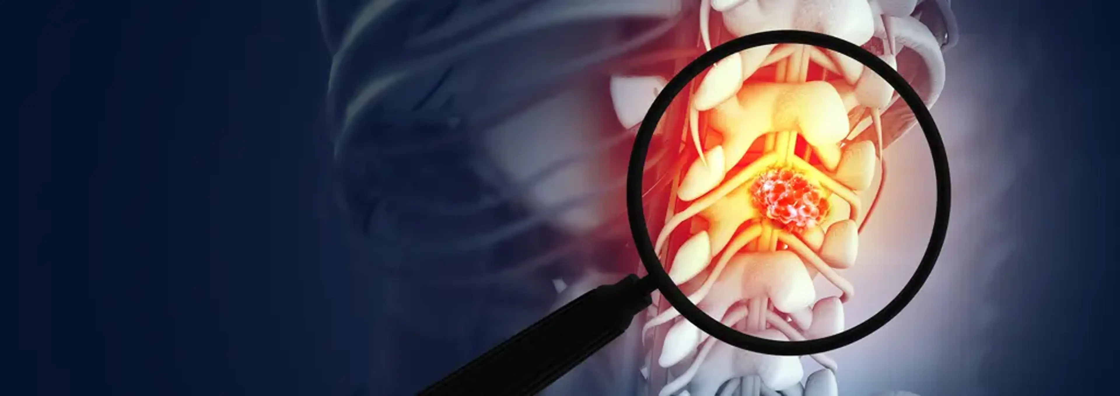
Introduction
introduction:-the-pursuit-of-natural-radianceBefore diving into stem cells, it’s important to understand why joint injuries — especially cartilage or deep joint damage — are such a difficult problem:
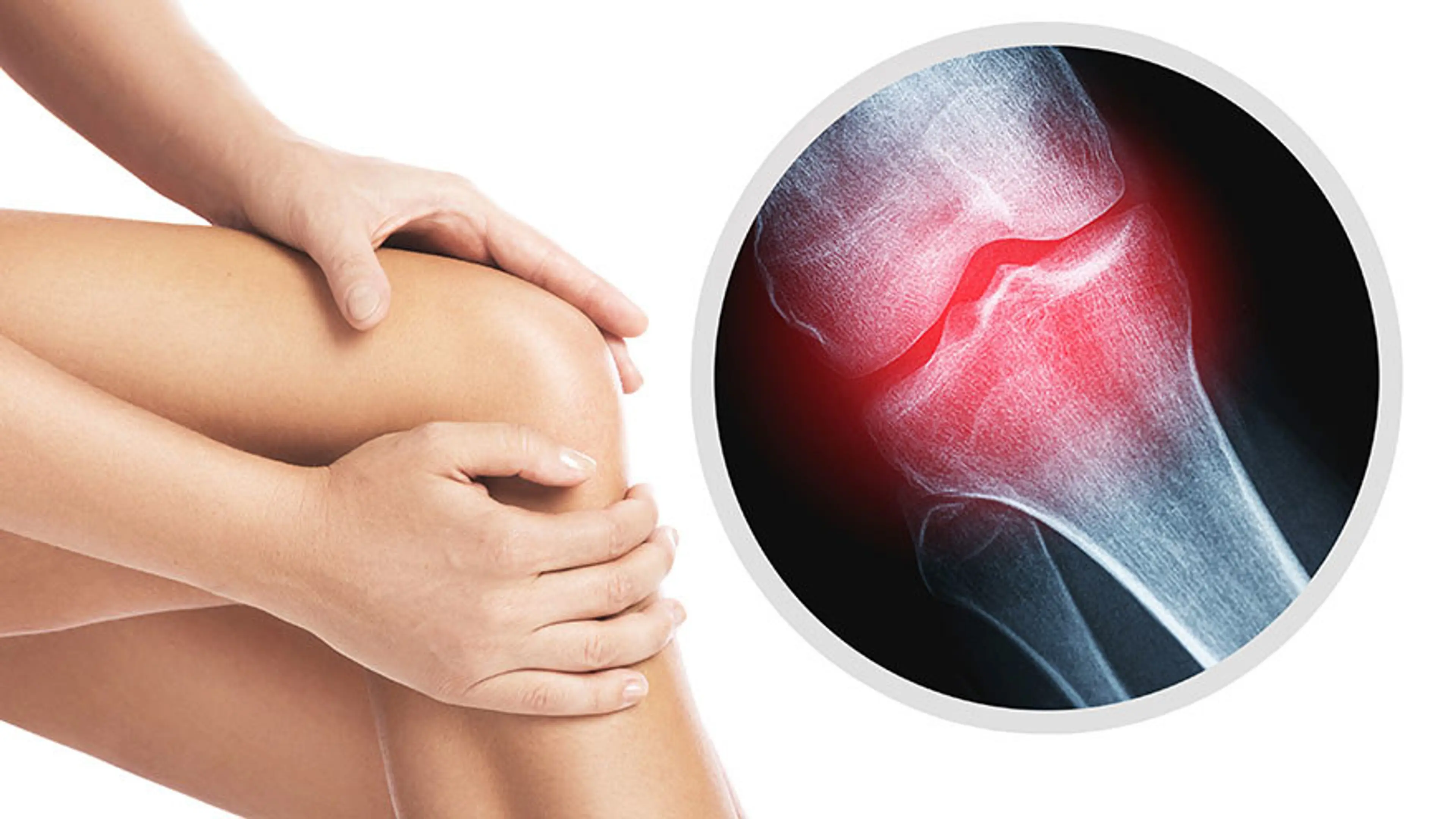
Cartilage has poor intrinsic repair capacity. Articular cartilage (the smooth, “slippery” tissue that cushions bones in a joint) is avascular (no blood vessels), and chondrocytes are relatively sparse. Once damaged, cartilage tends to degrade further or become replaced by inferior scar-like tissue.
Progressive degeneration risks “downward spiral.” Rough surfaces → more friction → inflammation → further cartilage breakdown → bone changes → pain & dysfunction.
Mechanical loading is unforgiving. Joints bear weight, move, twist, and absorb shocks continuously. Any repair must withstand mechanical stress, not just exist biologically.
Inflammation and catabolic environment. In osteoarthritis or chronic injury, the joint environment is hostile: inflammatory cytokines (IL‑1, TNF, MMPs) break down cartilage, inhibit repair, and create oxidative stress.
Mismatch between repair tissue and native cartilage. Even if some repair occurs (e.g. via microfracture techniques), the new tissue often becomes fibrocartilage — less resilient, less durable than hyaline cartilage.
So the key is: how to introduce or stimulate a repair that is biomechanically strong, durable, and compatible with the joint environment. Stem cell–based strategies seek to solve precisely that gap.
What Types of Stem Cells Are Used (or Proposed) in Joint Repair
what-types-of-stem-cells-are-used-(or-proposed)-in-joint-repairIn clinical and translational research, the primary stem cell type used for joint repair is mesenchymal stem / stromal cells (MSCs). Some salient features:
MSCs can be harvested from sources such as bone marrow, adipose (fat) tissue, synovium, umbilical cord, and placenta.
They are multipotent — i.e. under proper signaling, they can differentiate into cartilage (chondrocytes), bone (osteocytes), or other connective tissues.
Even more important than direct differentiation is their paracrine / trophic effect: MSCs secrete growth factors, cytokines, extracellular vesicles (exosomes), and signals that modulate inflammation, recruit resident progenitor cells, and orchestrate repair.
Because they exert immunomodulatory effects, MSCs are relatively safe for allogeneic (donor‑derived) use under controlled conditions.
Other more experimental or adjunct cell types include autologous chondrocytes / chondrons (cartilage cells taken from the patient) or hybrid approaches combining MSCs + chondrons. For example, the IMPACT / RECLAIM strategies use MSCs together with autologous chondrons to stimulate repair.
Additionally, perinatal stem cells (from amniotic fluid or umbilical tissue) are being studied for their high proliferation rates and anti-inflammatory profiles, though these are less commonly used in commercial or clinical settings.
Mechanisms: How Stem Cells Actually Help Heal Joints
mechanisms:-how-stem-cells-actually-help-heal-jointsStem cell therapy in joints is not magic — it works via several interacting biological mechanisms. Here's a breakdown:
1. Differentiation into Cartilage-Like Cells (Chondrogenesis)
1.-differentiation-into-cartilage-like-cells-(chondrogenesis)Under the right cues (growth factors, scaffold environment, low oxygen tension), MSCs can differentiate into chondrocytes, depositing cartilage-specific extracellular matrix (e.g. collagen II, aggrecan). This gives a “building block” for new cartilage repair.
However, in many human joint applications, full differentiation into mature cartilage is partial or limited — achieving a hyaline‑like repair is a central challenge.
2. Paracrine / Secretome Effects (Trophic Support)
2.-paracrine-secretome-effects-(trophic-support)One of the most powerful actions of MSCs is via the molecules they secrete:
Growth factors (e.g. TGF‑β, IGFs, BMPs) that promote cell proliferation, matrix deposition, and chondrogenesis
Anti-inflammatory cytokines and modulators (e.g. IL‑10, PGE2) that help quiet the joint’s inflammatory milieu
Extracellular vesicles / exosomes that deliver microRNAs, peptides, and proteins to local cells
Angiogenic/vasculature-modulating signals (though cartilage is avascular, modulation of subchondral bone is relevant)
These signals “coach” the existing tissue environment: suppress excessive inflammation, reduce catabolic enzymes (MMPs), recruit resident progenitor / stem cells, and support cell survival in a harsh environment.
3. Immunomodulation & Inflammation Control
3.-immunomodulation-and-inflammation-controlBecause degenerative joints are often inflamed, turning off that “hostile environment” is critical. MSCs can:
Reduce pro-inflammatory signaling (TNF, IL‑1)
Inhibit further cartilage breakdown by suppressing matrix-degrading enzymes
Modulate synovial inflammation
Reduce oxidative stress and apoptosis of resident chondrocytes
This “resetting” of the joint environment is a key enabler of regeneration.
4. Recruitment of Endogenous Progenitor / Stem Cells
4.-recruitment-of-endogenous-progenitor-stem-cellsEven if transplanted MSCs do not survive indefinitely, their signals can attract and activate the patient's own progenitor cells (e.g. from bone marrow, synovium, periosteum) to participate in repair. In effect, MSCs act as conductors rather than the sole builders.
5. Remodeling of Subchondral Bone & Interface Integration
5.-remodeling-of-subchondral-bone-and-interface-integrationJoint damage often involves subchondral bone (the bone just below cartilage). MSCs can help:
Stabilize or regenerate subchondral bone
Promote better integration of new cartilage with underlying bone
Prevent cysts, sclerosis, or bone irregularities
This integration is essential — if the cartilage “floats” over a weak bone base, it will fail under mechanical load.
Clinical Evidence: What Human Trials & Meta-Analyses Show
clinical-evidence:-what-human-trials-and-meta-analyses-showIt’s crucial to distinguish hopeful research from rigorously proven therapies. Here’s a summary of what human clinical data suggest so far.
Meta-Analyses and Systematic Reviews
meta-analyses-and-systematic-reviewsA recent meta‑analysis of randomized controlled trials (RCTs) evaluated intra-articular MSC injections for knee osteoarthritis. The findings:
MSCs significantly improved pain and function (e.g. WOMAC, VAS) at 6 and 12 months compared to control.
High-dose MSCs and adipose-derived MSCs showed more pronounced effects in subgroup analyses
No significant increase in adverse events compared to controls, implying reasonable safety in the treatment contexts studied
This suggests that MSCs are not merely safe, but clinically beneficial — at least in moderate to moderately severe osteoarthritis without co‑interventions.
Another review of cell‑based therapy in musculoskeletal disease underscores the ability of MSCs to support cartilage and bone repair, especially when combined with scaffolds or biomaterials.
A deeper review of MSC trials in OA notes that while many show symptomatic improvements and even imaging evidence of cartilage change, full-thickness, durable hyaline cartilage regeneration is still rare in human studies.
Recent Controlled / Blinded Studies
recent-controlled-blinded-studiesA triple-blind clinical trial tested adipose-derived MSCs (ADMSCs) versus placebo in knee OA patients. The MSC group had better pain relief (VAS) and even measurable increases in cartilage thickness on MRI, albeit with mixed effects on broader functional scores (KOOS).
A Phase 3 trial is ongoing (or completed) investigating JOINTSTEM® — autologous adipose-derived MSC injections into knees with K&L grade 3 osteoarthritis. The goal is to regenerate cartilage and improve joint function.
At specialized centers (e.g. Mayo Clinic), newer hybrid surgical + regenerative methods are being trialed. For instance, RECLAIM / IMPACT approaches combine autologous cartilage fragments (chondrons) + allogeneic MSCs in a single-stage implantation, aiming to harness both structural and signaling functions. Early results show sustained pain relief and functional gains.
Putting It All Together: How Stem Cell Therapy Can Heal Severe Joint Damage
putting-it-all-together:-how-stem-cell-therapy-can-heal-severe-joint-damageLet me walk you through a conceptual “workflow” of how stem cell therapy may be applied in a severe joint case — e.g., a patient with partial-thickness cartilage loss, subchondral bone changes, pain, and functional limitation.
Patient & lesion evaluation
Determine extent and depth of cartilage damage (MRI, arthroscopy)
Assess joint alignment, mechanical loading, and contributing factors (malalignment, ligament instability, meniscus status)
Screen systemic health, inflammation, metabolic factors
Stem cell harvesting / procurement
Autologous (from the patient): e.g. bone marrow aspiration or adipose tissue liposuction
Allogeneic: pre-prepared MSC bank (less invasive, “off-the-shelf”)
Cell preparation & “priming”
Culture-expansion, characterization, quality control
Preconditioning / priming (e.g. with growth factors, mechanical loading, hypoxia) to boost chondrogenic potential
Optionally combining with scaffold biomaterials (3D matrix, hydrogels, meshes) or other cell types
Delivery into the joint / lesion site
Intra-articular injection: simple, minimally invasive, but dispersion and retention are challenges
Targeted implantation / surgical approach: precise placement into cartilage defect, often using scaffolds or fixatives
Hybrid approaches: e.g. debriding the lesion first (microfracture, drilling) then injecting/implanting MSCs to “boost” repair
Post-procedure environment support
Optimize mechanical loading (controlled rehab, offloading)
Anti-inflammatory support, nutrition, biologic co-factors
Possibly adjunct therapies: low-intensity ultrasound, platelet-rich plasma, growth factor injections, exosome boosters
Monitoring & follow-up
Clinical assessments: pain, range of motion, function
Imaging (MRI, arthroscopy) to assess cartilage thickness/regrowth
Adjustments in rehabilitation based on early response
Long-term integration & remodeling
The goal is that new cartilage (or cartilage-like tissue) integrates with existing cartilage and bone, matures, and withstands mechanical stresses over years
Ongoing remodeling, chondrogenesis, and maintenance under functional loads
Why Seoul Yes Hospital (or a Specialist Center) Matters in This Context
why-seoul-yes-hospital-(or-a-specialist-center)-matters-in-this-contextGiven this landscape, a clinic like Seoul Yes Hospital (with expertise in regenerative therapies, multidisciplinary teams, and infrastructure for controlled cell-based protocols) has particular advantages:
Integrated care: stem cell therapy must be combined with orthopedic, physical‑rehab, pain management, and imaging expertise
Quality control & safety: rigorous protocols, sterility, cell characterization, and regulatory compliance
Tailoring therapies: choosing the right cell source, delivery method, priming, biomaterials, and rehabilitation plan for each patient
Follow-up & feedback loops: longitudinal monitoring, incorporation of new innovations (exosomes, scaffold materials, mechanical adjuncts)
Patient education: ensuring realistic expectations, understanding risks, and emphasizing that regeneration is a process, not an instant fix
In other words, the success of stem cell therapy for joints depends not just on “the cells” but on the entire ecosystem of care. A specialized center maximizes that ecosystem.
Realistic Expectations & Advice for Patients Considering Stem Cell Joint Repair
realistic-expectations-and-advice-for-patients-considering-stem-cell-joint-repairIf you or someone you know is exploring stem cell therapy for a damaged joint, here’s what to keep in mind:
Start with a full evaluation — imaging, alignment, joint stability, and comorbidities (e.g. metabolic disease, obesity).
Best results are in moderate damage, not “end-stage destruction.” Regenerative therapy works better when residual structure and biological potential remain.
Ask detailed questions:
What cell source is used (autologous vs donor)?
How many cells (dose)?
How are they processed / characterized?
What delivery method (injection vs surgical implantation)?
What scaffolds, biomaterials, or adjuncts (e.g. growth factors, exosomes) are involved?
What is the monitoring and post-op rehab protocol?
Understand that improvement is gradual. Most patients experience incremental gains over months, not overnight “regeneration.”
Be wary of unproven “stem cell clinics.” Always confirm that the treatment is part of a regulated clinical trial or under institutional oversight.
Pair with good joint care fundamentals: weight control, alignment correction, physical therapy, anti-inflammatory strategies, nutrition, and joint offloading when needed.
Ask about long-term follow-up and published outcomes — ideally peer-reviewed data rather than anecdotes.
In Summary
in-summaryStem cell–based therapies represent one of the most exciting frontiers in joint repair: harnessing biological regeneration rather than just replacing or patching. While not yet flawless or universally effective, advances in MSC biology, scaffold materials, delivery systems, and clinical protocols are steadily pushing outcomes forward.
At a center with strong regenerative medicine expertise — like Seoul Yes Hospital — patients have the best chance of benefiting from this evolving science: carefully selected, meticulously programmed, and closely monitored. If joint pain and cartilage damage are limiting your life, exploring a stem cell–augmented regenerative plan may well be the turning point toward reclaiming function.